
During a whole abdomen scan, a technician applies a special gel to the patient's abdomen to ensure good contact between the ultrasound probe and the skin. The probe, or transducer, is then moved over the abdomen, emitting high-frequency sound waves that bounce off internal structures. These echoes are captured and converted into real-time images displayed on a monitor. The procedure is generally painless and takes about 30 to 60 minutes.
info@pranavmedhospital.com
+7845578465
Whole Abdomen
Whole Abdomen
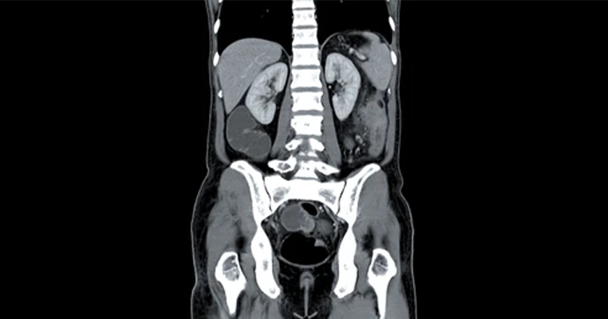
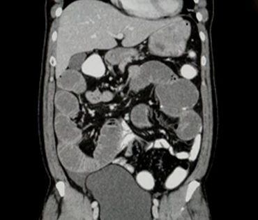
A whole abdomen scan, often referred to as an abdominal ultrasound, is a non-invasive imaging test used to evaluate the organs and structures within the abdominal cavity. This scan helps diagnose a variety of conditions such as liver disease, kidney stones, gallbladder issues, and abdominal tumors. It is also useful for assessing the size, shape, and position of organs and detecting any abnormalities, such as cysts or tumors.
How the Procedure is Performed
Indications for the Scan
-
Persistent abdominal pain or discomfort
-
Abnormal liver function tests
-
Suspected kidney stones or urinary tract infections
-
Gallbladder disease or gallstones
-
Monitoring of known abdominal conditions, such as tumors or cysts
-
Preoperative assessment before abdominal surgery
Benefits and Considerations
The benefits of a whole abdomen scan include its non-invasive nature, lack of ionizing radiation, and ability to provide detailed images of internal organs. It helps in early detection and diagnosis, which can be crucial for effective treatment. However, while the scan is generally safe, certain factors such as obesity or excessive gas can sometimes affect image clarity. It's important for patients to follow any preparatory instructions provided by their healthcare provider to ensure optimal results